1. Team Introduction:
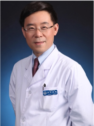
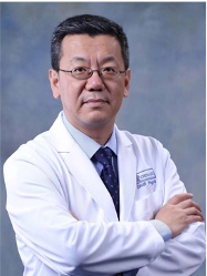
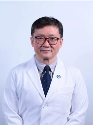
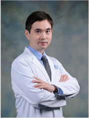
The Neurosurgical Center for Stereotactic Treatment and Functional Disorders is a professional group featuring epilepsy, trigeminal neuralgia, hemifacial spasm, microvascular decompression, and biopsy of the hard-to-diagnose diseases of the central nervous system. It brings together world-renowned specialists in Neurosurgery to design the most effective treatment for each patient's condition. The team leader of the group is Professor Guoguang Zhao. The leading experts are Professor Guoguang Zhao, Professor Yongzhi Shan, and Professor Yaming Wang.
After the China International Neuroscience Institute (CHINA-INI) building was put into use in 2019, the total number of beds in the Neurosurgical Center for Stereotactic Treatment and Functional Disorders has increased significantly. Nowadays, our center has 40 beds and 9 EEG monitoring devices, located on the third floor of the CHINA-INI building (Neurosurgery Ward INI3).
In terms of hardware, our center is equipped with an operating microscope, neuro-navigation system, robotic system, stereotactic EEG and supporting radiofrequency thermocoagulation system, intraoperative EEG and cortical stimulation system, evoked potential electrophysiological monitoring system, video EEG, magnetoencephalogram, transcranial magnetic stimulation system, neuropsychological and cognitive evaluation system.
In the past few years, the annual operation volume of our team was about 600 units/group. Four new technologies and businesses have been employed. Team members received 8.26 million yuan in various project funds, with two municipal talent projects/titles.
"The best way to predict the future is to create it." Since 2019, as the starting year, two national natural projects have been put into research status. We also completed the task of "Key R&D by the Municipal Science and Technology Commission" and successfully passed the final acceptance. In September 2019, our team was awarded the "National Neurosurgery Robot Application Demonstration Project Training Base" by the National Health Commission and the Ministry of Industry and Information Technology. Professor Guoguang Zhao is the chairperson of the "National Neurosurgery Robot Expert Steering Committee." Professor Yongzhi Shan serves as the secretary-general and the standing committee of the "National Neurosurgery Robot Expert Steering Commit." Our team takes the lead to promote the diagnosis and treatment of domestic neurosurgery robots.
2. Multidisciplinary discussion team:
◆ The multidisciplinary evaluation expert group of epilepsy
The constituent members include Professor Guoguang Zhao and Professor Yongzhi Shan of the Neurosurgery Department, Professor Yuping Wang and Professor Liankun Ren of the Neurology Department, and other Well-known experts and scholars at home and abroad such as Professor Liping Zhang of the Pediatrics Department. The multidisciplinary cooperation of Neurosurgery, Pediatrics, and Neuroimaging has become our center's characteristic diagnosis and treatment model.
◆ The multidisciplinary discussion expert group on stereotactic biopsy of intractable nervous system diseases
The constituent members include Professor Guoguan Zhao, Professor Yongzhi Shan, and Professor Yaming Wang of the Neurosurgery Department; Professor Cunjiang Li of the Neurology Department; Professor Dehong Lu, Professor Yueshan Piao of the Neuropathology Department; and Professor Zhilian Zhao of the Neuroimaging Department.
3. What We Treat
◆ Hypothalamic hamartoma
A hamartoma is a benign (non-cancerous) growth made up of an abnormal mixture of cells and tissues. A hypothalamic hamartoma is a tumor-like formation within the hypothalamus, the area at the base of the brain that controls the production and release of hormones by the pituitary gland. These hormones regulate a wide array of bodily functions, from growth and metabolism to sexual development and the body's reaction to stress.
Due to its deep location and adjacent to the critical structures, craniotomy is extremely risky, the postoperative complication rate is high, and the effect of epilepsy treatment is unreliable. Our center adopts radiofrequency damage treatment after stereotactic electrode implantation. This technology clarifies the location of epileptic foci and destroys them in a minimally invasive manner without craniotomy. (According to statistics, more than 60% of patients have no seizures after surgery. Only a few of patients require secondary damage)
◆ Hippocampal sclerosis (medial temporal lobe epilepsy)
Hippocampal sclerosis is the most common drug-refractory epilepsy. It usually manifests as abdominal discomfort, fear, Déjà vu, and other auras. Sometimes it appears dumbfounded, slapped, and fumbled, which may cause body twitches. Magnetic resonance can find apparent hardening of one side of the hippocampus. The anterior temporal lobe resection has a complete control rate of more than 80%. On this basis, our center also carries out the stereotactic electrode damage to achieve minimally invasive treatment without craniotomy. The chances of staying seizure-free after one year of treatment are similar to traditional anterior temporal lobectomy, but it has better protection for patients' cognition and visual field.
◆ MR negative drug-resistant epilepsy
Drug-resistant epilepsy is when medicines cannot control seizures. Because there is no visible lesion in the brain tissues of the patient, the abnormal areas can only be inferred indirectly through clinical manifestations, EEG, and brain metabolism. After confirmed by intracranial electrode EEG, further radio-frequency damage or surgical resection can be performed. Relying on the solid support of the neurology and neuro-electrophysiology department, our center has accumulated rich theoretical and practical experiences in the surgical treatment of MR negative medically refractory epilepsy.
◆ Trigeminal neuralgia
Trigeminal neuralgia is a chronic pain condition that affects the trigeminal nerve, which carries sensation from your face to your brain. It is commonly seen in middle-aged and older adults. Carbamazepine is usually effective, but the pain will gradually worsen after the drug is taken for long-term. Studies have found that most trigeminal neuralgia is caused by the tortuous blood vessels in the brain that compress the root of the trigeminal nerve. Therefore, the disease can be cured by microvascular decompression surgery without taking medications every day.
◆ Hemifacial spasm
Hemifacial spasm is a neuromuscular disorder characterized by frequent involuntary contractions (spasms) of the muscles on one side (hemi-) of the face (facial). The disorder occurs in both men and women, although it more frequently affects middle-aged or older women. Studies have found that the disease is usually caused by intracranial blood vessels compressing the root of the facial nerve. Facial nerve microvascular decompression currently is the treatment with the highest cure rate and the best curative effect.
◆ Glossopharyngeal neuralgia
Glossopharyngeal neuralgia consists of recurring attacks of severe pain in the back of the throat, the area near the tonsils, the back of the tongue, part of the ear, and/or the area under the back of the jaw. The pain is due to a malfunction of the ninth cranial nerve (glossopharyngeal nerve). Studies have found that most glossopharyngeal neuralgias are caused by the tortuous blood vessels in the brain that oppress the glossopharyngeal nerve roots. Therefore, the disease can be cured by microvascular decompression surgery without taking medicines every day.
◆ Nervous system tumors or complex diseases
Some intracranial tumors (such as lymphoma) or rare and difficult neurological diseases require histopathological examinations to be diagnosed and further treated. Our center will cooperate with the Department of Neurology, Imaging department, and Pathology department to collect intracranial specimens accurately and safely in diagnosis and treatment. Intracranial cystic space-occupying lesions such as craniopharyngiomas, brain abscesses, and intracranial hematomas can be diagnosed by biopsy with stereotactic technology. And we implant drainage tubes, shunt tubes, Omaya capsules, etc. for drainage or drug delivery.
4. Treatment
◆ Stereo electroencephalography (SEEG)
Stereotactic electroencephalography (SEEG) utilizes localized, penetrating depth electrodes to measure electrophysiological brain activity. It is most commonly used in the identification of epileptogenic zones in cases of refractory epilepsy. Our center has adopted this technology beforehand in China and accumulates many practical experiences in the early stage. At present, we have multiple sets of robot-assisted stereotactic operating systems. A dedicated team is responsible for the implementation of this technology. We have reached the highest level in terms of safety and accuracy internationally. Our center's lever of evaluation, location, and surgical results of epileptic foci are similar to those of the world-class centers.
◆ Radiofrequency thermocoagulation treatment of intracranial epileptic foci
If the foci are multiple or difficult to remove completely, we can treat epilepsy by destroying the epileptic foci by intracranial radiofrequency thermocoagulation.
It should be noted that currently only hypothalamic hamartoma, medial temporal lobe epilepsy, and paraventricular gray matter heterotopia are expected to achieve long-term satisfactory results through this method. In the treatment of other types of epilepsy, it is still a palliative or experimental treatment. Patients need specific guidance from their doctors.
◆ Precise resection of epileptic foci
After the epileptic foci have been identified through non-invasive or invasive examination, they can be accurately removed by microsurgery.
With neuro-navigation equipment, the area to be removed can be marked in advance. The important intracranial structures such as functional areas, conduction bundles, blood vessels, etc. also can be marked. During the operation, the epileptogenic foci can be accurately positioned and safely removed without causing damages to other functions. In some operations involving the critical functional areas such as exercise and language, the intraoperative wake-up technique can be used to accurately remove the epileptic foci while preserving the patient's brain functions to the greatest extent.
◆ Multiple lobectomies and hemisphere resection
Some particular types of diseases, such as Rasmussen encephalitis, Sturge-weber syndrome, hemispheric softening, and drug-refractory epilepsy caused by hemispheric giant gyrus, may require extensive surgical resection. Some patients even need to be removed the entire hemisphere. Using advanced technologies such as imaging and physiological evaluation, we can accurately predict the brain's compensation before surgery and complete these operations at the least cost.
◆ Neuromodulation surgery
For drug-refractory epilepsy that is difficult to cure by radical surgery, vagus nerve stimulation is a good option. This kind of operation has high safety, no damage to brain tissue and nerves. According to the statistics, more than half of the patients have improved to varying degrees after treatments.
◆ Transcranial magnetic stimulation
Using external magnetic signals to stimulate brain tissue, this medical technology affects the electrophysiological process of brain tissues and then produces the effect of locating epileptic foci or treating epilepsy.
◆ Microvascular decompression
Microvascular decompression (MVD) is a surgery to relieve abnormal compression of a cranial nerve causing trigeminal neuralgia, glossopharyngeal neuralgia, or hemifacial spasm, glossopharyngeal neuralgia and other diseases. Through a 5cm skin incision inside the hairline, the compressed nerves and blood vessels are microscopically separated without damaging the brain tissues and cranial nerves. According to the statistics, the surgical cure rate is about 80%.
◆ Stereotactic biopsy
A stereotactic biopsy is a surgical procedure where a thin needle is inserted into the brain by a neurosurgeon to extract a small piece of tissue to examine under a microscope to help diagnose and treat nervous system tumors or complex cases. It can be used for diagnostic sampling of lymphoma, glioma, inflammatory granuloma, and other complex diseases.
◆ Stereotactic implantation
It is mainly used for cystic intracranial lesions, drainage, or local medication, including drainage of brain abscess, drainage of various cysts, drainage and local medication for craniopharyngioma, local medication for glioma, etc.
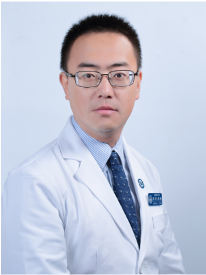
You can find professional doctors and experts about this center here for your further consultation
Any use of this site constitutes your agreement to the Terms and Conditions and Privacy Policy linked below.
A single copy of these materials may be reprinted for noncommercial personal use only. "China-INI," "chinaini.org" are trademarks of China International Neuroscience Institute.
© 2008-2021 China International Neuroscience Institute (China-INI). All rights reserved.