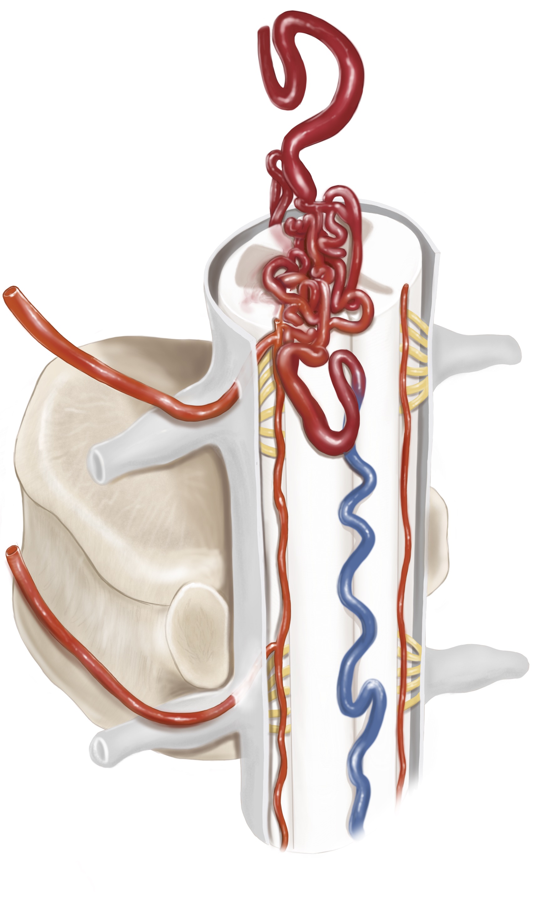
1. Overview
Normally, as blood is pumped away from the heart, it travels through the aorta to arteries, and then the capillary beds. After oxygen is removed from the blood during the capillary bed, the blood flows to the the veins back to the heart. Arteriovenous malformation is a collection of unmatured (dysplastic) dilated blood vessels (neither arteries nor veins) wherein arterial blood flows directly into draining veins without the normal interposed capillary beds. AVMs are usually congenital, tend to enlarge and progress with age. AVMs appear grossly as a “tangle”of vessels, often with a fairly well-circumscribed center (the nidus) and a draining “red veins” (usually on the surface) containing oxygenated blood under higher pressure than normal veins.
2. Presentation
◆ Seizures: Seizures are waves of abnormal electrical activity in the brain. They can make you pass out, or move or behave strangely.
◆ Symptoms of hemorrhage: headaches, passing out, nausea and vomiting and a stiff neck.
◆ Symptoms of a stroke–A stroke is when a part of the brain is damaged because of a problem with blood flow. A stroke can cause:
① A person's face to look uneven or droop to 1 side
② Weakness or numbness in 1 or both arms–For example, 1 arm might drift down if a person tries to hold both arms out.
③ Trouble speaking, or speech that sounds strange
④ Sudden, severe headache
3. Evaluation
Various imaging modalities provide information that are additive when analyzed in combination. Definitive diagnosis and aspects of treatment planning are obtained with catheter angiography. In catheter angiography, a thin plastic tube, called a catheter, is inserted into an artery through a small incision in the skin. Once the catheter is guided to the area being examined, a contrast material is injected through the tube and images are captured using a small dose of ionizing radiation (x-rays). Cross-section based modalities (CT, CTA, MRI, MRA) provide important information about adjacent brain that is not obtained with catheter angiography.
4. Treatment
AVMs that cause bleeding usually need treatment. The different treatments for an AVM include:
◆ Surgery to remove the AVM
◆ Radiosurgery – This is not surgery. It involves getting radiation (high doses of X-rays) in the area of the AVM. Over time, the radiation makes the AVM less likely to bleed.
◆ Embolization–This is a procedure done during cerebral angiography. The doctor puts a material into the blood vessel that brings blood to the AVM. The material blocks off the blood vessel. Sometimes this procedure works on its own. Other times, doctors do surgery or radiosurgery after it.
You can find professional doctors and experts about this disease here for your further consultation and treatment.


Any use of this site constitutes your agreement to the Terms and Conditions and Privacy Policy linked below.
A single copy of these materials may be reprinted for noncommercial personal use only. "China-INI," "chinaini.org" are trademarks of China International Neuroscience Institute.
© 2008-2021 China International Neuroscience Institute (China-INI). All rights reserved.