1. What is Gamma Kinfe?
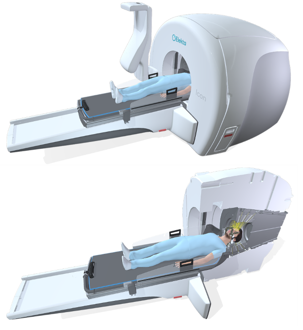
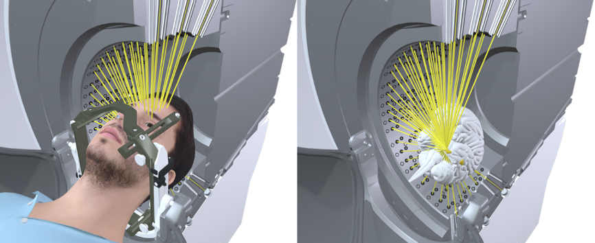
Gamma Knife is a type of radiation therapy, also known as stereotactic radiosurgery, and is one of the most advanced and non-invasive options available today for the treatment of intracranial tumors and other lesions. The main structure of Gamma Knife is 192 60Co radioactive sources (60Co is one of the radioactive isotopes of the element cobalt) arranged in a hemispherical pattern. These sources produce gamma rays, which are focused precisely through image guidance, so that gamma rays from different directions are concentrated on intracranial lesions, forming a high-energy radioactive target area with the exact shape of the lesion, thus killing tumor cells while hardly affecting the surrounding normal tissues. The effect of radiation treatment is just like the removal of surgical knife, therefore called gamma knife.


Xuanwu Hospital Capital Medical University is one of the first bases of neurosurgery and cradle of neurosurgeon’s cultivation in China, founded in 1958 by Professor Yicheng Zhao, who is known as "the father of Chinese neurosurgery", and has developed into a prestigious medical, teaching and research center at China and abroad. It is the national center for neurological disorders, the first batch of national clinical specialties, the first batch of neurosurgery specialist training base and China's neurosurgery training base for foreign aid.
Since the first gamma knife was successfully developed in Sweden in 1967 and first introduced into China in the 1990s, the gamma knife has been continuously updated and has served in China for more than 30 years, and is a mature and widely recognized treatment method. Globally, more than 80,000 patients are treated with Gamma Knife each year. Gamma Knife has the highest degree of focus precision and the smallest error among existing types of radiation therapy, and is therefore called the "gold standard" of stereotactic radiosurgery.
Xuanwu Hospital of Capital Medical University has been carrying out gamma knife radiosurgery since 2006 under the leadership of its current dean, Prof. Guoguang Zhao. The Gamma Knife Center of Xuanwu Hospital Capital Medical University has equipped the most advanced Leksell® Gamma Knife Icon™, with an average radiation precision of 0.15 mm and a comprehensive treatment precision within 0.5 mm, which can treat brain metastases, vascular malformations, and other neurological disorders in the deep brain area without causing other injuries.
2. What we treat: the indications of Gamma Knife Radiosurgery
(1) Intracranial metastases, hemangiopericytomas, perivascular cell tumors, lymphomas, and certain type of gliomas.
(2) Intracranial vascular malformations: arteriovenous malformations, arteriovenous fistulas, cavernous sinus cavernous malformations.
(3) Small and medium-sized benign intracranial tumors: vestibular schwannoma, trigeminal schwannoma, pituitary adenoma, meningioma, craniopharyngioma, etc.
(4) Functional diseases: trigeminal neuralgia, tremor palsy, etc.
(5) Residual or recurrent tumors after craniotomy.
(6) Tumors of skull base and intracranial-extracranial communication: intraorbital tumors, chordoma, tumor in the jugular foramen area, metastases, and tumors of the upper cervical spine.
(7) Otolaryngology, ophthalmic tumors: tumors of the sinus or paranasal sinus cavity, intraorbital and fundus tumors.
3. The advantages of Gamma Knife Radiosurgery
(1) Low risk: safe and effective, with much lower risk than traditional neurosurgery, especially suitable for deep brain lesions that are difficult to safely reach with surgery.
(2) No incision: No general anesthesia is required, no surgical incision is made, and there is usually no or only slight discomfort after treatment.
(3) Less damage: minimal damage to the normal tissue surrounding the lesion.
(4) Short time: short hospitalization time, only 1-2 days, and can treat several lesions in different areas at one time.
(5) Less costly: the cost is lower than that of traditional neurosurgery, and it has been included in the national medical insurance.
4. Treatment procedure
The Gamma Knife treatment process is divided into 4 steps: install the head frame, image positioning, developing a treatment plan, and Gamma Knife treatment. Before the treatment, your doctor will explain the procedure and the potential risk and ask you and your family to sign an informed consent.
(1) Installation of the head frame: one of the prerequisites to ensure the accuracy of Gamma Knife treatment is the head frame fixation. A stereotactic head frame made of a lightweight alloy is fixed to your head with four head pins, and the doctor and nurse will pre-inject local anesthetic into the pins.

(2) Image positioning: Once the head frame is in place, your doctor will take you to the imaging department for a magnetic resonance imaging (MRI) exam. Patients with cerebral arteriovenous malformations will also undergo a cerebral angiogram. During these exams, your head frame will be secured to the exam bed to ensure accurate image acquisition.

(3) Treatment planning: Once the imaging is complete, your doctor will design the treatment plan based on these data. Each treatment plan must be discussed and approved by a multidisciplinary team of specialists before implemented.

(4) Gamma Knife treatment: Once the treatment plan is ready, you will be transported to the treatment center. The positioning head frame will be fixed to the Gamma Knife treatment bed to ensure precise treatment. At the start of the treatment, the treatment bed will move smoothly to bring your head into the treatment chamber. The length of treatment varies depending on the size and number of lesions, usually within one hour. The medical staff will continuously monitor your treatment in the control room, and you can communicate with the medical staff at any time through the video and voice system in the treatment room.

At the end of the treatment, the medical staff will remove the head frame and bandage the fixed area, and then send you back to your room to rest.
Gamma Knife treatment is painless, with no burning sensation, noise, or other discomfort. During the treatment, you can listen to soothing music or close your eyes to relax.
It takes some time for the results to show after the Gamma Knife treatment. Gamma Knife stereotactic radiosurgery works by causing the tumor or diseased tissue to stop growing and gradually shrink. In general, brain metastases can be seen to shrink significantly in 1 month and the related symptoms improve, brain arteriovenous malformations can be seen to shrink in about 6 months and basically completely occluded in about 2-3 years, and meningiomas can be seen to shrink partially in about 6 months. These effects may take weeks or even years to gradually appear until the effects are maximized, so you will need to stay in touch with your doctor and make a follow-up appointment with him or her at a scheduled time.
4. Unique-Features of treatment in Gamma Knife Center of Xuanwu Hospital Capital Medical University
According to the indications for Gamma Knife treatment and the types of diseases admitted to form a series of special treatment diseases
(1) Overall neurosurgical solutions for brain metastases and MDT discussion
According to the specific situation of brain metastases, different solutions or combinations of solutions are selected.
The comprehensive neurosurgical solution for brain metastases
Gamma knife alone: single and multiple tumors
Stereotactic aspiration + Gamma knife treatment: cystic brain metastases
Stereotactic aspiration biopsy + Gamma knife treatment: intracranial first symptom onset, clear primary focus
Gamma knife first, then craniotomy: multiple brain metastases with cranial hypertension but no brain herniation
Craniotomy first, then gamma knife: single/multiple metastases with brain herniation, tumor cavity irradiation
MDT multidisciplinary discussion
Internal medicine: combined gamma knife + targeted drugs/immunotherapy for brain metastases
Radiotherapy department: Gamma knife + whole brain radiotherapy WBRT combined treatment of brain metastases
Out-of-hospital cooperation: RCT research, academic conference
(2) Individualized treatment of cerebral arteriovenous malformation
Localization methods
Combined DSA+MRI localization of vascular malformations
Treatment modality
Targeted embolization + gamma knife for high-grade AVM
Gamma knife + interventional embolization for simultaneous treatment
Radiosurgery volume splitting and dose splitting treatment for complex AVMs
(3) Individualized treatment of craniopharyngioma
Including postoperative recurrent craniopharyngioma gamma knife treatment and cystic craniopharyngioma stereotactic aspiration (Ommaya capsule implantation) + gamma knife treatment
(4) Special patients admitted according to the characteristics of the neurosurgery subspecialty group
a) Individualized treatment of cerebral arteriovenous malformations in young children as young as 3 years old
b) Gamma knife treatment for elderly patients with trigeminal neuralgia
c) Gamma knife treatment for C2-C3 chordoma and fibroblastoma
You can find professional doctors and experts about this center here for your further consultation


Any use of this site constitutes your agreement to the Terms and Conditions and Privacy Policy linked below.
A single copy of these materials may be reprinted for noncommercial personal use only. "China-INI," "chinaini.org" are trademarks of China International Neuroscience Institute.
© 2008-2021 China International Neuroscience Institute (China-INI). All rights reserved.