Department of Neurosurgery, Xuanwu Hospital 2024-01-28 20:11 Beijing
The brain is the most sensitive organ to blood oxygen content, and the oxygen content in brain tissue directly affects brain metabolism and functional integrity. Abnormal oxygen metabolism is also closely related to neurological lesions caused by craniocerebral trauma, ischemic-hypoxic encephalopathy, epilepsy, and tumors, etc. In addition to dramatic fluctuations in electrophysiology, it will also be accompanied by disturbances in oxygen metabolism especially in the course of epileptic seizures. Therefore, it is of great significance to observe and analyze the law of oxygen metabolism in the brain of epilepsy patients to improve the clinical diagnosis and treatment of epilepsy. Compared with the blood oxygen information obtained by near-infrared spectroscopy, functional magnetic resonance and other techniques, tissue oxygen partial pressure is more closely related to local neural activities and metabolic processes. How to monitor the oxygen partial pressure of brain tissue in real time, accurately, conveniently and in situ, and analyze its interaction with neural activities is also key issue in the basic research of brain science and clinical diagnosis and treatment of brain diseases. Recently, in order to broaden the modal selection in epilepsy diagnosis and treatment and epileptic brain network research, and realize continuous monitoring of brain oxygen metabolism in epileptic patients, time monitoring of oxygen partial pressure in the deep brain tissue of freely moving animals, the research group of Professor Zhao Guoguang, Department of Neurosurgery, Xuanwu Hospital, together with the research group of Sheng Xing, Department of Electronic Engineering, Tsinghua University, and the research group of Ding He, School of Optoelectronics, Beijing Institute of Technology, developed an implantable micro-photoelectric probe, which can be used to realize wireless, continuous, real-time monitoring of oxygen partial pressure in the deep brain tissue of freely moving animals.

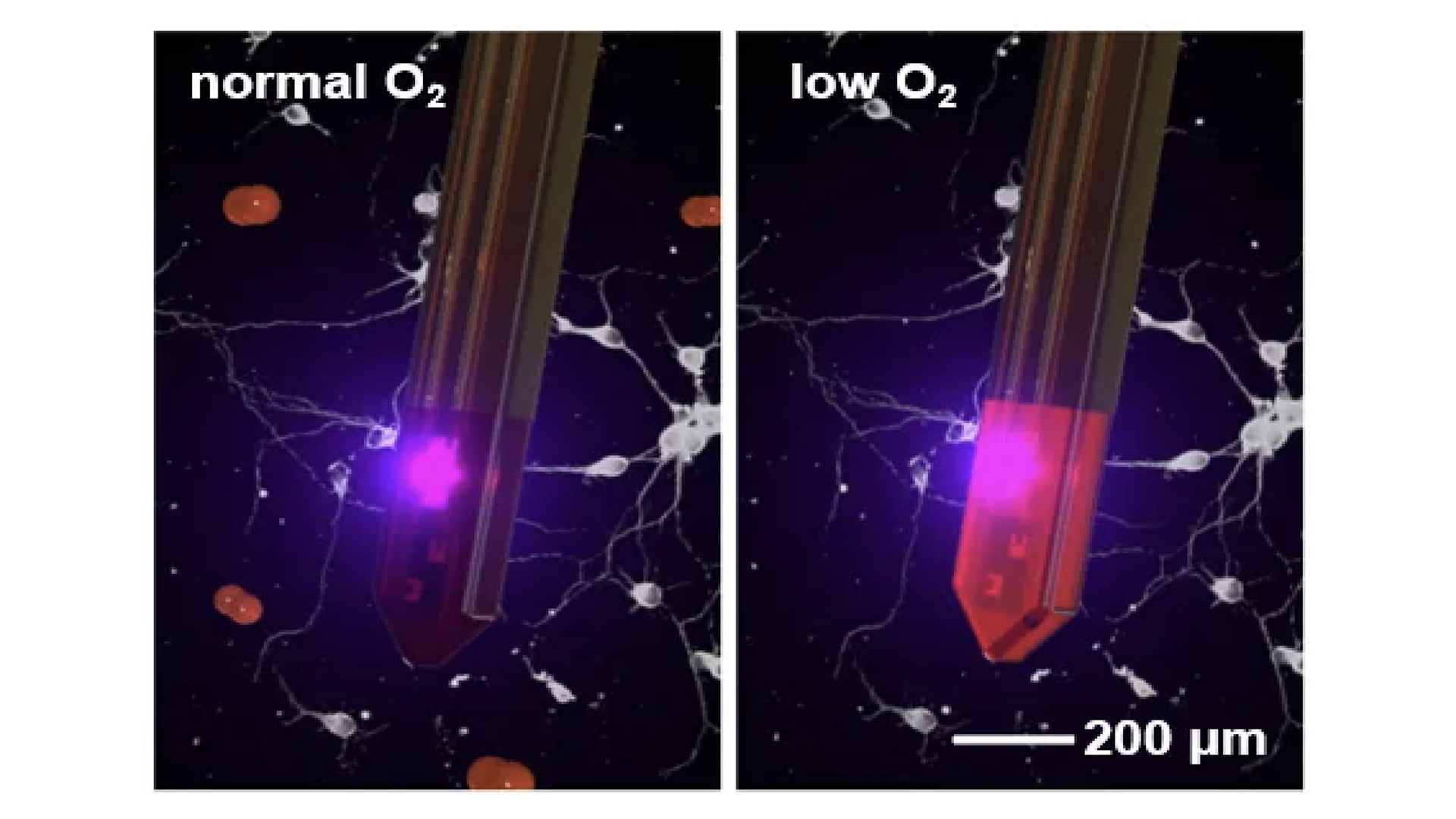
Figure 1 Working schematic diagram of the oxygen partial pressure detection probe in brain tissue.
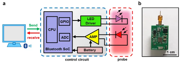
In this work, a highly integrated miniature implantable photoelectric probe is created by designing and preparing miniature thin-film light-emitting diodes (LEDs) and photodetectors, and combining them with oxygen-sensitive phosphorescent coatings to realize oxygen partial pressure detection by using the phosphorescent bursting effect of oxygen on platinum-based metal compounds. In addition, the research team developed a miniature flexible circuit for wireless energy and signal transmission, together with the oxygen partial pressure detection probe, to realize a fully implantable photoelectric oxygen partial pressure sensing system.

Figure 2 Wireless circuit design
In the in vitro characterization test, the phosphorescent signal intensity measured by the oxygen partial pressure detection probe decreases with the increase of oxygen partial pressure, and the probe response time is less than 1 second, and it has a high oxygen selectivity. Rodents (mice, etc.) were used as models to achieve wireless dynamic monitoring of oxygen partial pressure in the deep brain region of animals through minimally invasive implantation, and to explore the changes of oxygen partial pressure of brain tissues in the case of changing the concentration of inhalation oxygen, short-time carotid artery clamping, and anesthesia. In addition, in the model of epilepsy induced by electrical stimulation of the hippocampal brain region in mice, the significant oxygen deprivation and oxygen partial pressure fluctuation of different brain regions, such as the hippocampus and the cortex, were measured after epileptic discharges. Combined with electrophysiological recordings, the changes of neural electrical activity and oxygen partial pressure of local tissues were monitored synchronously during epilepsy, and the roles and its mechanism of oxygen in the process of abnormal neural activity, as well as the coupling law between the local neural activity and the metabolism of oxygen were explored. This wireless, real-time, in situ, accurate and dynamic oxygen partial pressure monitoring technology provides an effective tool for in-depth exploration of the relationship between oxygen metabolism and neurological activities and brain disease states.

Figure 3 Changes in oxygen content of animal brain tissue were measured by wireless oxygen partial pressure detection system under the condition of changing inhalation oxygen and carotid artery ligation

Figure 4 Dynamic changes of oxygen content in hippocampus were detected by wireless oxygen partial pressure detection system in epileptic discharge state of mice.
The research results were recently published in the journal Nature Photonics (Nature Photonics), entitled A Wireless Optoelectronic Probe to Monitor Tissue Oxygenation in Deep Brain Tissue). Professor Guoguang Zhao, Department of Electronics, Professor Sheng Xing, the Department of Electronics, Tsinghua University, Tsinghua-IDG/McGovern Institute of Brain Science, and Professor Ding He, Beijing Institute of Technology, are the co-corresponding authors; and the co-first authors are Cai Xue, PhD student the Department of Electronics, Tsinghua University, Wei Penghu, associate professor of Neurosurgery, Xuanwu Hospital; and Liu Quanlei, PhD student. The remaining co-authors are from Beijing Institute of Technology and Minzu University of China. This research was supported by the Beijing Natural Science Foundation, the National Natural Science Foundation of China, and the Ministry of Science and Technology.
Any use of this site constitutes your agreement to the Terms and Conditions and Privacy Policy linked below.
A single copy of these materials may be reprinted for noncommercial personal use only. "China-INI," "chinaini.org" are trademarks of China International Neuroscience Institute.
© 2008-2021 China International Neuroscience Institute (China-INI). All rights reserved.