Department of Neurosurgery, Xuanwu Hospital 2024-02-06 19:30 Beijing
The “Tuesday Review of Surgery” is a surgical review session for Department of Neurosurgery in Xuanwu Hospital, which has been a long-standing tradition and brand activity of our department. A resident neurosurgeon will introduce each carefully selected surgery with clinical information and play the edited surgical video with key surgical procedures. Then the lead neurosurgeon will summarize the surgical highlights and the surgical team will take questions from the audience around the whole department. In this rigorous and open discussion, fellow neurosurgeons analyze the pros and cons of each case, and learn from each other’s experiences to improve themselves. In order to encourage the continuous pursuit of the perfection of knowledge and techniques, the department upgraded the surgical review session to “Skills of Xuanwu Neurosurgery-New Technique / Complex Surgery / Treatment of critical case” competition. The audience will score each case according to the difficulty, presentation, and discussion. The winning cases will be shared on publicizing platforms such as WeChat public account, YouTube, TikTok and so on. We welcome comments and suggestions from domestic and foreign colleagues!
Difficult cases of "Skills of Xuanwu Neurosurgery" in 2024
--Skull Base and Brain Tumor Center
Difficult Cases
Epidural abrasion for resection of medial meningioma in the anterior bed eminence of the sphenoid crest
Operators: Bai Jie, Xiao Xinru
Medical history
Female, 50 years old Complaint: headache and dizziness for half a year, vision loss in the right eye for 2 months. Past history: healthy Examination: visual acuity 0.5 on the right and 0.8 on the left, weakness of eyelid elevation in the right eye, narrowing of the eye fissure.
Preoperative imaging examination
Cranial MRI enhanced
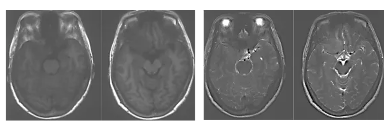
1. MRI T1 and T2 images showed a solid tumor in the medial region of the right sphenoid crest with long T1 and short T2 signals, and peritumoral edema was not obvious.
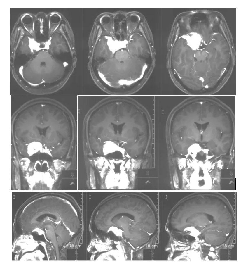
2. Enhanced axial, coronal and sagittal positions showed that the tumor strengthened uniformly with a broad base, mainly located in the medial part of the right sphenoid crest, the base of middle cranial fossa, the saddle diaphragm, encroaching on the cavernous sinus bilaterally, and the internal carotid artery, the middle cerebral artery and the anterior cerebral artery and their perforating blood vessels were encircled by the tumor.
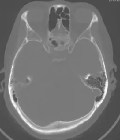
CT bone window of the skull base
No significant enlargement of the right optic canal
Diagnosis.
Right medial sphenoid crest meningioma
Preoperative discussion
The medial region of the right sphenoid crest is occupied, with the main symptoms of decreased vision in the right eye and narrowing of the right ocular fissure. The tumor had clear boundaries and uniform enhancement. The preoperative diagnosis was benign meningioma, and the indications for surgery was relatively clear. The basal extent of the tumor was wide, involving the right anterior bedspace, saddle node, saddle diaphragm, and bilateral cavernous sinuses. The internal carotid artery, middle cerebral artery and anterior cerebral artery and their perforating vessels were encircled by the tumor. Overall, the tumor was divided into two parts, one in the subdural and one in the bilateral cavernous sinuses. Cavernous sinus meningiomas are generally hard, and surgical resection is more damaging to the cranial nerves, especially the motor nerve, and is not easy to remove completely. Therefore, cavernous sinus tumors are not removed for observation or adjuvant treatment, and only subdural tumors are removed in this surgery. Subdural invasion of the saddle diaphragm region tumor management is a difficult surgical point, and the patient's vision loss, optic nerve canal opening is also necessary. Considering the above, abrasion of the anterior bed protrusion and opening of the optic nerve canal can decompress the optic nerve, increase the optic nerve mobility, facilitate the resection of the tumor in the optic nerve canal, and increase the exposure of the tumor in the saddle-diaphragm region. Intraoperative separation of the internal carotid artery, middle cerebral artery and their branches from the tumor is another difficult part of the surgery, especially the protection of some of the perforating vessels is the key to avoid postoperative neurological dysfunction.
Treatment strategy
Right frontotemporal approach epidural abrasion of the medial sphenoid crest tumor resection in the anterior bed.
Auxiliary means: electrophysiologic tests, including SEP and MEP
Surgical position: supine position with the head tilted 30 degrees to the left
Intraoperative
After the head position was fixed by a metal head frame and the sterile sheet was routinely disinfected, the frontotemporal arc incision was designed to be long. The scalp was incised, the inter fascial approach was used, the temporalis muscle was separated to expose the skull, and the frontotemporal bone flap was shaped by milling cutter. The dura mater of the anterior and middle cranial fossa was separated, the orbital meningeal ligament was severed, the anterior clinoid process and the upper wall of the optic canal were removed with highspeed drill, and the optic canal was opened.
The dura mater was cut open in an arc, the lateral fissure was separated, and the M2 branch was exposed. The tumor base was located in the anterior bed process, the saddle node, the base of the middle cranial fossa, and it grew upward toward the saddle, and the internal carotid artery and its branches were encircled by the tumor, and the optic nerve was shifted to the left by the tumor extrusion and thinning. The tumor was first dissected from the base of the anterior bed process and the base of the middle cranial fossa, and the anterior temporal vein was protected from draining into the sphenoid sinus, and the tumor was decompressed by resecting part of the tumor in pieces. Then the tumor was dissected at the base of the saddle node, the sickle ligament and part of the optic nerve sheath were incised, the tumor in the optic canal was excised, and the tumor was dissected at the base of the saddle diaphragm through the second and third interspaces. The tumor was separated from vascular adhesions along the middle cerebral artery to protect the branches of the internal carotid artery and the perforating vessels. After resecting most of the tumor in pieces, the cerebellar vermis was incised along the actinic nerve, and the tumor located at the edge of the cerebellar vermis was resected to protect the actinic nerve. The subdural tumor was completely resected under the microscope. The operation area was strictly hemostatic, the dura mater was sutured, the bone flap was repositioned and fixed, the scalp was sutured layer by layer, and the operation was finished. After removal of the metal head frame, the patient returned to the ICU with a tracheal tube. 12 hours later, the patient recovered from spontaneous breathing
After the tracheal intubation was removed, he could answer correctly and move his limbs voluntarily. the right eye fissure was the same as before the operation, and the visual acuity of the right eye was 2 ft and the left side was the same as before the operation.
Pathologic diagnosis
Atypical meningioma, WHO grade II.
Postoperative review
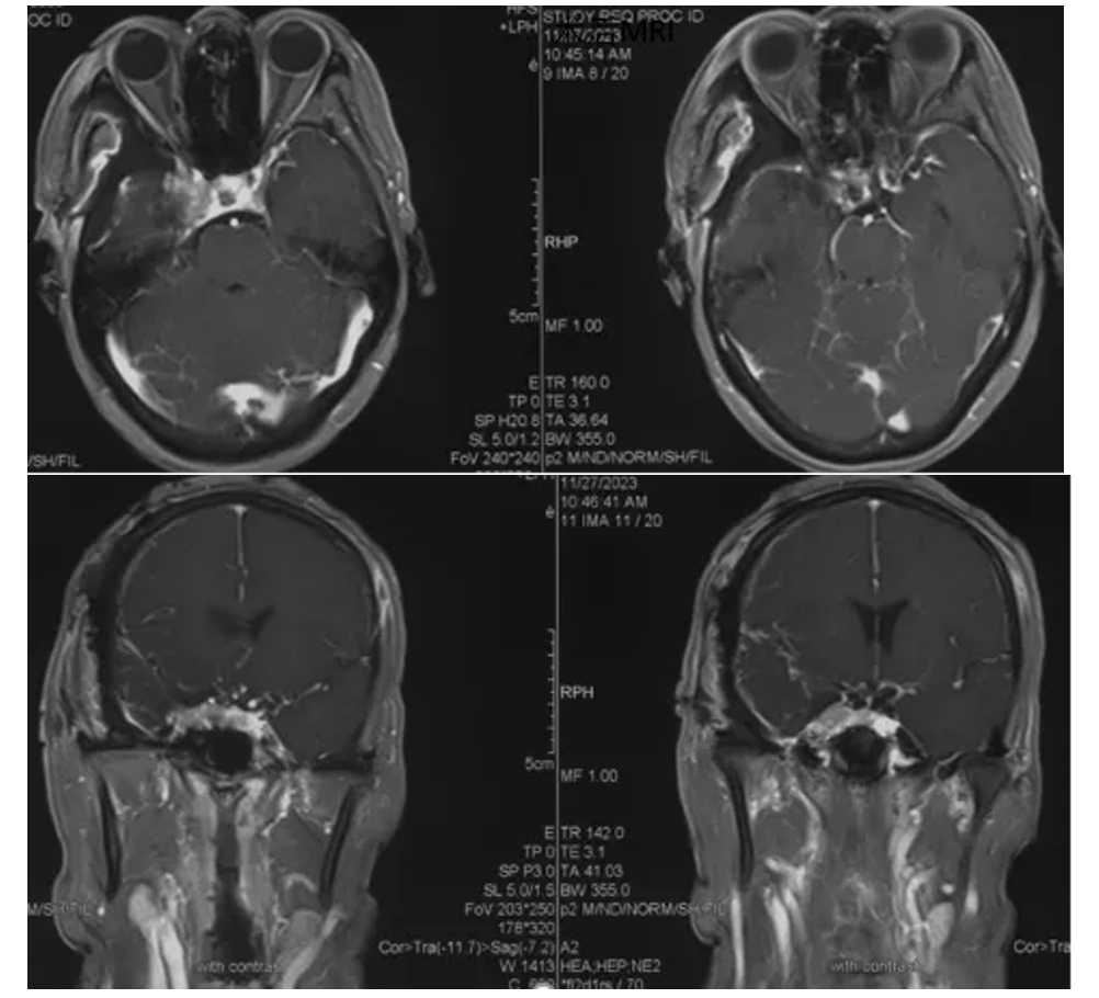
Postoperative MRI enhancement
Showing complete resection of the tumor.
Postoperative situation
Postoperative eye movement photo
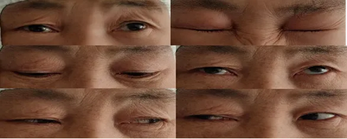
Postoperative limb movement
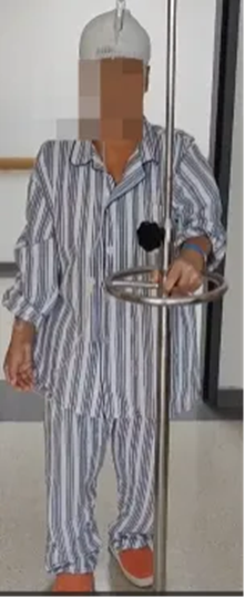
(Patient information is for professional discussion only with the consent of the patient and his family)
Operator's comment
Sphenoid crest meningioma often straddles on the sphenoid ridge, growing forward and above the anterior cranial fossa, growing backward and below the middle cranial fossa, starting from the anterior bed process on the inside, reaching the sphenoid point on the outside. The incidence of sphenoid ridge meningioma accounts for 13%-19% of intracranial meningioma. Medial pterostriatal meningioma refers to the meningioma originating from the anterior bed process or the medial pterostriatal wing, accounting for 50%-60% of pterostriatal crest meningiomas. The location of the medial sphenoid ridge meningiomas are deep, easily invade the optic nerve, internal carotid artery and its branches, cavernous sinus and other important anatomical structures, and partly invade orbital growths, so they are difficult to be operated with poor prognosis, and are one of the difficult problems in neurosurgery. Currently, surgical resection is still its main treatment modality, but total surgical resection is difficult, with poor prognosis and high recurrence rate. The resection rate and prognosis are related to the relationship between the tumor than the neighboring, texture, size, and peritumoral edema. If the tumor base is extensive, especially if it is accompanied by vision loss and the tumor grows toward the optic nerve canal, grinding off the anterior bed protrusion and opening the upper wall of the optic nerve canal is an effective means of complete resection of the tumor, and it can also increase the degree of freedom of the optic nerve, and the increase of the second gap space is conducive to the dissociation of the tumor located in the saddle region of the base and the tumor is completely resected. This surgical technique was adopted in this case and the subdural tumor was completely resected. Tumors in the cavernous sinus region can be selected for observation or other adjuvant treatments according to the pathological nature of the tumor after surgery.
The Department of Neurosurgery in Xuanwu Hospital has performed nearly 1,000 meningiomas a year. Our team annually completes a large number of meningiomas in skull base, deep, huge and recurrent every year, and has accumulated relatively rich clinical experience in the treatment of meningiomas. The treatment effect has been recognized by the majority of patients. We will do our best to help each patient to get rid of the pain and work as much as possible to help them return to normal life.
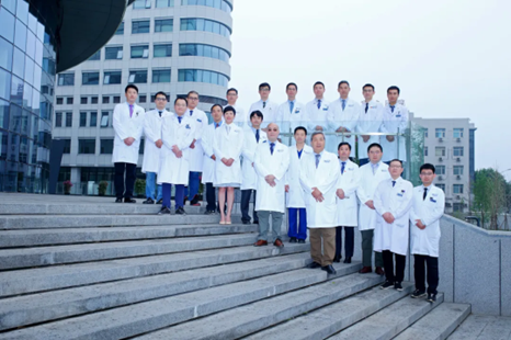
Group photo of Skull Base and Brain Tumor Center
Any use of this site constitutes your agreement to the Terms and Conditions and Privacy Policy linked below.
A single copy of these materials may be reprinted for noncommercial personal use only. "China-INI," "chinaini.org" are trademarks of China International Neuroscience Institute.
© 2008-2021 China International Neuroscience Institute (China-INI). All rights reserved.