The “Tuesday Review of Surgery” is a surgical review session of department of neurosurgery in Xuanwu Hospital and a long-standing tradition and brand activity. Each carefully selected surgery is presented by a resident neurosurgeon with clinical information and edited surgical video with key surgical procedures. The lead surgeon will summarize the surgical highlights and the surgical team will take questions from the audience from the whole department. In this rigorous and open discussion, fellow neurosurgeons analyze the merits and demerits of each case, learn from each other’s experiences in order to improve themselves. To encourage the continuous pursuit of the perfection of knowledge and techniques, the department upgraded the surgical review session to “Skills of Xuanwu Neurosurgery-New Technique / Complicated Surgery / Treatment of critical case” competition. The audience will score each case according to the difficulty, presentation, and discussion. The winning cases will be shared in publicizing platforms such as Wechat public account, Youtube, Tiktok and so on. We welcome the comments and suggestion from domestic and foreign colleagues!
Pediatric posterior fossa ependymoma resection
Summary of medical history
6-year-old boy, right handed, 16 kg
Intermittent nausea and vomiting for 2 months, combined with unsteady walking for 1 month.
Physical examination: ataxia; normal muscle tone and strength; no signs of cerebral nerve palsy
Pre-operative imaging
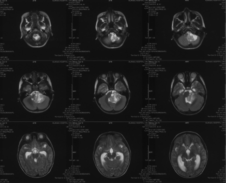
Preoperative imaging MRI-T2
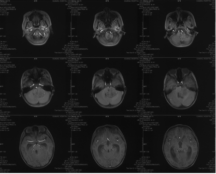
Preoperative imaging MRI-T1
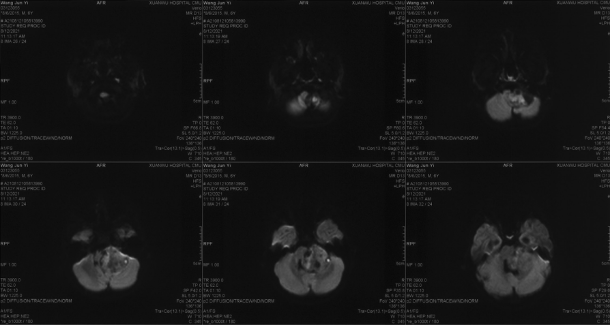
Preoperative MRI-DWI
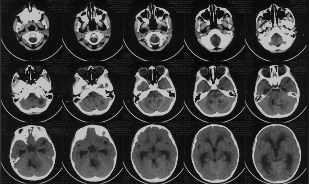
Preoperative CT
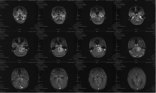
Preoperative enhanced MRI
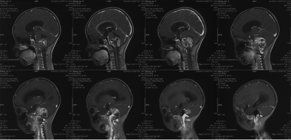
Preoperative enhanced MRI
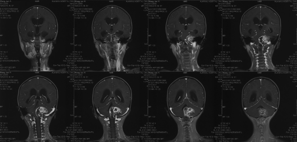
Preoperative enhanced MRI
Diagnosis and surgical strategy
Diagnosis: posterior fossa occupying lesion: ependymoma(?); obstructive hydrocephalus
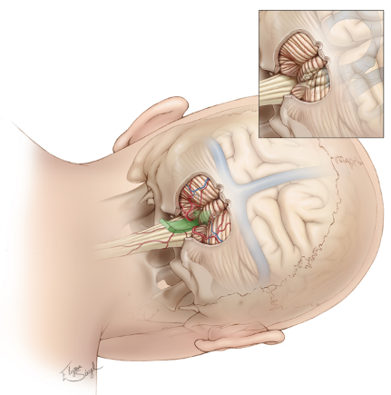
Surgical strategy: tumor resection via suboccipital midline approach, right lateral position
Postoperative pathological results: posterior cranial fossa ventricular meningioma, PFB group

Gross findings: Frozen for examination:
(Posterior cranial fossa occupancy) grayish gray-red unshaped tissue, 1 cm in diameter, medium quality, locally soft. Also examined:
(Posterior cranial fossa) grayish-brown abnormal tissue, size 3x2x0.8cm, medium
Pathological diagnosis:
(Posterior cranial fossa) mesenchymal ventricular meningioma with nuclear schizophrenia and focal necrosis.

Immunohistochemical results:
(#3) EMA (paranuclear punctate +), D2-40 (+), GFAP (+), 0lig-2 (-),vimentin (+), Ki-67 (hot spot region 25% +), L1CAM (-), Cyclin D1 (partial +), H3K27me3 (+), P53 (partial +).
PFB ventricular meningioma
Molecular subgroup risk stratification is better than histopathological grading
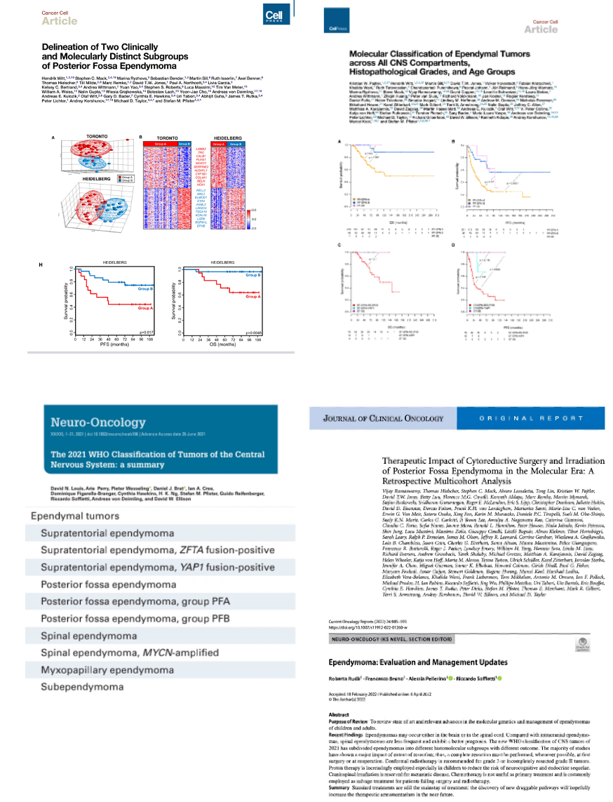
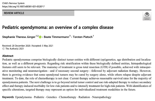
The current data from literature suggest that some patients with EPN_PFB can be cured by surgery alone after GTR. A large proportion of EPN_PFB patients can still be successfully treated with re-surgery and delayed radiotherapy even if they relapse after initial cessation of radiotherapy."
"To date, data from prospective clinical studies for different molecular subgroups are not available. Some of the hypotheses generated in retrospective translational studies need to be tested in clinical trials."
1-year postoperative imaging and follow-up
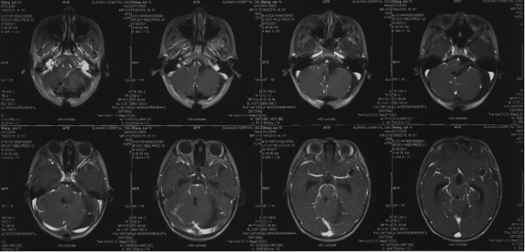
Postoperative enhanced MRI
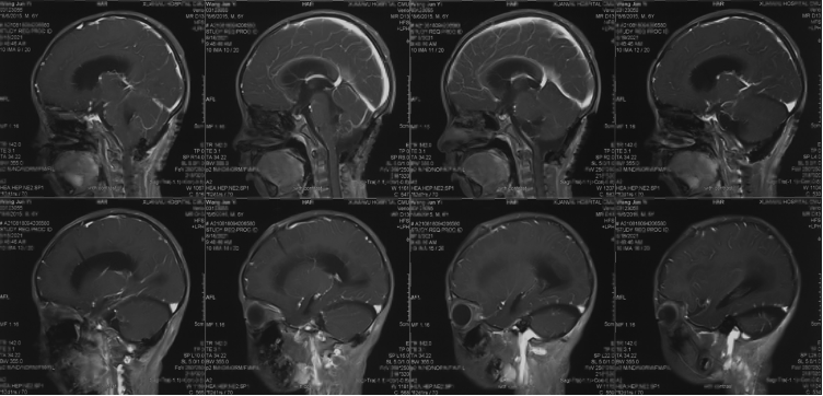
Postoperative enhanced MRI
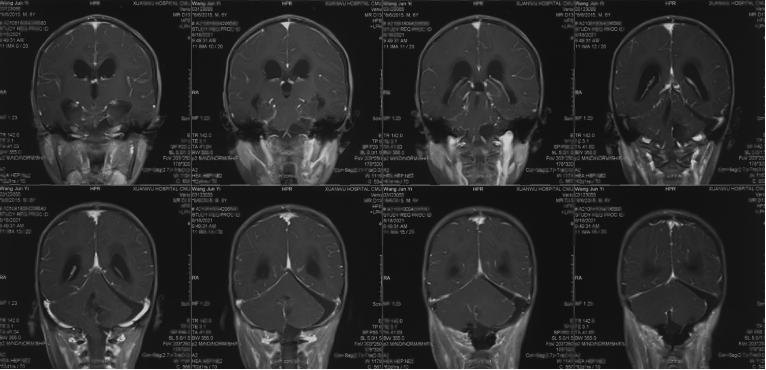
Postoperative enhanced MRI
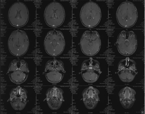
Postoperative follow-up 1 year enhanced MR
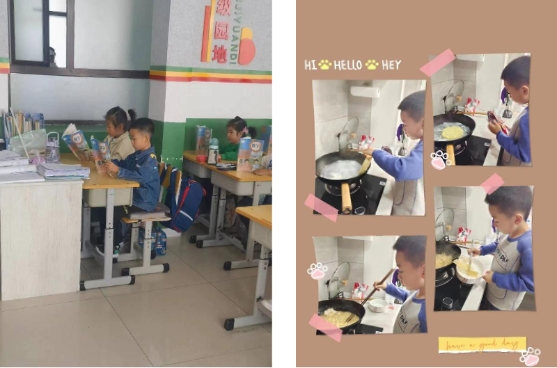
Picture of playing and reading 1 year after surgery
Summary
The ability to completely excise the tumor remains the primary determinant of ependymoma (regardless of supratentorial/infratentorial, molecular typing). For supratentorial ependymoma growing toward the lateral foramen of the quadrigeminal ventricle, the focus needs to be on protecting the lower group of cranial nerves, delineating the lateral interface of the medulla oblongata, combining blunt and sharp dissection, and making full use of the arachnoid interface.
For EPN_PFB with GTR, observation or reduced dose of radiotherapy is an option.
The vast majority of children with hydrocephalus secondary to posterior fossa tumors do not require shunt surgery.
Surgeon’s remark
Children are not shrunken versions of adults, and pediatric nervous system tumors are also vastly different from adult nervous system tumors. Pediatric neurosurgery is not just about surgery, but should also focus on the progress of related disciplines and the long-term prognosis of the children.
Any use of this site constitutes your agreement to the Terms and Conditions and Privacy Policy linked below.
A single copy of these materials may be reprinted for noncommercial personal use only. "China-INI," "chinaini.org" are trademarks of China International Neuroscience Institute.
© 2008-2021 China International Neuroscience Institute (China-INI). All rights reserved.