Cavernous sinus hemangioma (CSH) is a rare extra-axial highly vascular neoplasm that accounts for 2% to 3% of all cavernous sinus tumors. They are benign, well-encapsulated neoplasms arising within the confines of the cavernous sinus. Hemangiomas most often present in middle-aged patients and are more common in women.
CSH may present with headache or various neurological features, including visual diminution, diplopia or facial hypesthesia. Although a number of authors have described their extensive experience with the surgical treatment of cavernous sinus lesions with good clinical results, these uncommon lesions remain a challenge for the neurosurgeon, as there are high mortality and morbidity rates associated with uncontrollable and massive hemorrhage during surgery. Current treatment modalities include microsurgical resection, embolization, fractionated radiation therapy and stereotactic radiosurgery (SRS). The optimal management strategy is still a matter of controversy. Complete resection of CSH is potentially curative but may be complicated by profuse intra-operative bleeding and new cranial neuropathies. Gamma Knife radiosurgery (GKRS; Elekta AB, Stockholm, Sweden) has been performed for high-risk or residual CSH.
Cavernous sinus hemangiomas are not true neoplasms and are better described as vascular malformations. In the central nervous system, they are encountered most commonly in the brain parenchyma. Extra axial hemangiomas are rare and can occur in the cavernous sinus or cerebellopontine angles and have clinical behavior and imaging features different from their intra-axial counterparts. Extra-axial hemangiomas occur more commonly in middle-aged women. Microscopically, these lesions consist of numerous dilated vascular channels lined by endothelial cells.
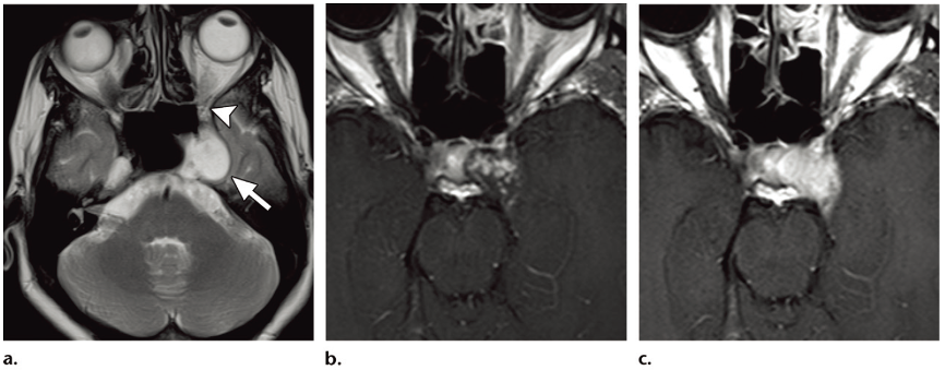
At MRI, cavernous hemangiomas are characterized by high signal intensity on T2-weighted and FLAIR MR images. Dynamic contrast enhanced T1-weighted MR images show characteristic progressive “fill-in” of contrast agent, with intense homogeneous enhancement on late gadolinium-enhanced MR images. Hemangiomas encasing the cavernous segment of the ICA usually do not cause luminal narrowing of the ICA, in contrast to meningiomas, which tend to cause luminal narrowing of the ICA. Scintigraphic imaging after administration of 99mTc pertechnetate–labeled red blood cells demonstrates accumulation of tracer within the lesion, a finding that is said to be specific for making the diagnosis of cavernous hemangioma. Digital subtraction angiography may show hypertrophic branches from the ICA and the external carotid artery, as well as variable vascular blush. Establishing the diagnosis of cavernous hemangioma before surgery is of paramount importance, because these tumors are notorious for causing profuse and often uncontrollable intraoperative hemorrhage.

Figure: Cavernous hemangioma in a 40-year-old woman with decreased vision in the left eye that had been present for 6 months. No other clinically important cranial nerve deficits were seen. (a) Axial T2-weighted MR image shows a homogeneous hyperintense mass (arrow) in the left cavernous sinus and the left trigeminal cave (Meckel cave). The superior orbital fissure (arrowhead) is not involved. (b) Axial T1-weighted MR image obtained 2 minutes after contrast agent injection shows central enhancement within the lesion. (c) Axial T1-weighted MR image obtained 9 minutes after contrast agent injection shows complete fill-in of contrast agent, with homogeneous enhancement. (Note that the gadolinium-enhanced images in b and c have a different axial orientation, compared with the axial T2-weighted image in a.)
You can find professional doctors and experts about this disease here for your further consultation and treatment.
Any use of this site constitutes your agreement to the Terms and Conditions and Privacy Policy linked below.
A single copy of these materials may be reprinted for noncommercial personal use only. "China-INI," "chinaini.org" are trademarks of China International Neuroscience Institute.
© 2008-2021 China International Neuroscience Institute (China-INI). All rights reserved.